Dental Implants After Severe Lower-Jaw Bone Loss: What the Jaw Keeps, and the Options That Hold Up Over Ten Years
People with a very thin lower jaw are often told there is nothing to put an implant into. Usually that describes the back of the jaw, not the whole of it. The front of the lower jaw tends to keep usable bone long after the rest has gone, and the long-term trials in exactly these patients point to simpler solutions than most people expect.


Written and medically reviewed by Dr. Sam Jain, DMD, MS, and Dr. Arpana Gupta, DDS, MDS
Published
In this article
- 1Why the lower jaw loses so much, and what it keeps
- 2What "severe" means on a scan
- 3Implants in the front of the jaw holding a removable denture
- 4Fixed teeth on implants in the front of the jaw
- 5Short implants, grafts and moving the nerve
- 6Risks that are specific to a very thin lower jaw
- 7Choosing between fixed and removable when the jaw is thin
Why the lower jaw loses so much, and what it keeps
A jaw has two layers of bone. The alveolar bone is the part that exists to hold teeth; once the teeth are gone, it has no job and resorbs. The basal bone beneath it forms the body of the jaw and stays. A classic study of 300 skulls, which produced the classification surgeons still use, found that the shape of the basal bone in both jaws remains relatively stable while the alveolar bone changes markedly in height and width, and in a predictable pattern.
Dentures speed the loss in the back of the lower jaw because they press on bone that nothing else is loading. In a five-year study, the back of the lower jaw lost an average of 1.63 mm of height under conventional dentures, compared with 0.69 mm in patients whose lower denture was held by two implants. Over a mean of 10.5 years in another group of 82 patients with implant-retained lower dentures, the back of the jaw lost only 0.5 mm, a change the authors did not consider clinically relevant.
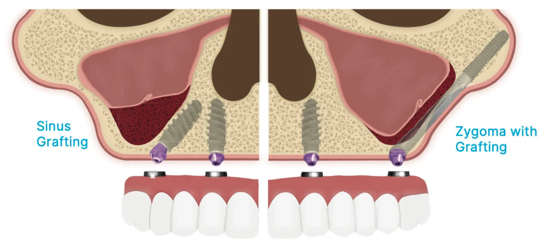
The practical consequence is geography. In the back of a severely resorbed lower jaw, the nerve canal may be only a few millimeters below the crest, so there is little room for an implant. In the front, between the two openings where the nerve leaves the bone, there is no main nerve canal in the way, and the basal bone often still offers enough height for implants. That is where most solutions start.
What "severe" means on a scan
The words patients hear, not enough bone, knife-edge ridge, pencil-thin jaw, describe different problems. A ridge that is tall but narrow needs a different answer from one that is wide but short, and a jaw that is short only at the back needs a different answer from one that is short everywhere. Only a 3D scan separates them: it measures the height from the crest to the nerve canal at each point, the width of the ridge, the position of the openings where the nerve exits, and the thickness of the jaw itself.
Your CBCT scan is taken free at the consultation, and it is read site by site rather than as a pass or fail for the whole mouth. Our article on whether a site can be made suitable for an implant explains the measurements; this one is about what can be done when those measurements are at the severe end.
Implants in the front of the jaw holding a removable denture
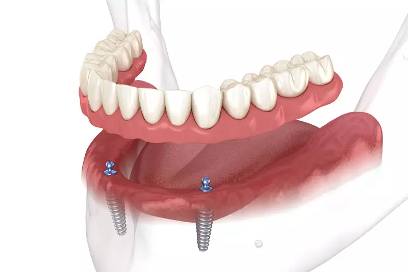
For people who have lost all their lower teeth, a denture that snaps onto two implants in the front of the jaw was proposed as the first-choice standard of care in the 2002 McGill consensus statement, and it remains a strong option when the jaw is very thin. The implants go where the bone still is, the denture stops moving, and the load on the back of the jaw falls, which is consistent with the slower bone loss described above.
The most useful evidence for severe cases comes from a randomized trial of 60 patients with extremely resorbed lower jaws, each given an implant-retained overdenture by one of three routes: a large transmandibular implant through the whole jaw, a bone graft from the hip followed by four implants, or four short implants placed without grafting. After ten years, implant survival was 76.3%, 88% and 98.8% respectively, and 30%, 5% and 0% of patients had needed further surgery. Satisfaction, psychological well-being and chewing improved significantly in all three groups, with no meaningful difference between them. Patients rated the hip graft the least favorable route because of its pain, discomfort and a surgical phase of at least six months. The authors concluded that short implants with an overdenture should be the first choice for these patients.
Our snap-in denture page explains how overdentures work day to day and how implant positions can be chosen so the plan can later be converted to fixed teeth.
Fixed teeth on implants in the front of the jaw
Fixed full-arch teeth in the lower jaw usually rely on the same front section of bone. In the All-on-4 approach, four implants are placed between the nerve openings, with the back two angled to spread their support toward the molars. In a study of 245 patients with implants placed in the front of the lower jaw to support fixed full-arch teeth, 99.2% of the bridges remained in service at up to ten years, and 94.8% of the implants met the study's success criteria. That study did not select patients for severe bone loss, so its numbers describe the approach rather than the most extreme jaws.
A fixed bridge on implants also appears to protect the bone behind the last implant. In 60 patients with lower fixed bridges on five or six implants, the height of the jaw measured behind the implants increased from 7.25 mm to 8.18 mm over four years, most of the gain in the first year of function. Whether four, six or eight implants are right for a given jaw depends on bone, bite force and the length of the bridge; our comparison of All-on-4, All-on-6 and All-on-8 explains the trade-offs, and All-on-4 here is one fixed, all-included fee per arch, while an arch that needs six or eight implants is quoted in writing.
Short implants, grafts and moving the nerve
When teeth are needed further back than the front section can support, the choices are shorter implants, rebuilding the bone, or moving the nerve. Our research review on short implants versus bone grafting goes through the randomized trials in the lower jaw in detail; in brief, short implants have generally matched grafted sites on survival with fewer complications, while extra-short implants under 8 mm call for more caution.
Grafting is not obsolete, and sometimes it is the right answer, especially when height is needed for appearance or when implants of any length cannot be placed safely. Moving the nerve aside to make room is a third option some surgeons use, with temporary sensory disturbance in 15% to 40% of cases in a 2025 meta-analysis; our article on numbness after implant surgery explains that trade-off and why distance from the nerve matters.
Risks that are specific to a very thin lower jaw
A severely resorbed lower jaw is weaker than a normal one. Fracture of the front of an atrophic jaw after implant placement is rare, but it is a recognized complication in the surgical literature, which is one reason implant size, number and position are planned against the thickness of the jaw on the scan, not just its height, and why a soft diet is followed while the implants heal.
The nerve is the other risk. In a thin jaw the openings where the nerve exits can sit close to the top of the ridge, and the nerve may loop forward inside the bone before it exits. Those structures are located on the CBCT scan before surgery, and the implants are placed with X-Guide or Navident EVO dynamic navigation, which shows the drill's position against the plan in real time.
Choosing between fixed and removable when the jaw is thin
Both can work in a severely resorbed lower jaw, and the evidence does not declare one better for everyone. A snap-in denture on implants needs fewer implants, is easier to clean, and its pink base can replace a large amount of lost bone and support the lip. Fixed teeth never come out, feel more like natural teeth, and need a steadier daily cleaning routine underneath. For some patients the bone decides; for many it is a choice about daily life, budget and appearance.
Because the planning, surgery and lab work happen in one building, both options are designed from the same scan and smile photographs, and the quote for each is itemized in writing. If you have been told elsewhere that your lower jaw is too thin for implants, a second opinion with a free 3D scan is a reasonable next step before accepting that answer.
Written and medically reviewed by Dr. Sam Jain, DMD, MS, and Dr. Arpana Gupta, DDS, MDS, who own this practice. Reviewed
This article is patient education, reviewed by Dr. Sam Jain, DMD, MS, and Dr. Arpana Gupta, DDS, MDS, and is not a substitute for an exam. Treatment recommendations are made only after an in-person consultation and 3D imaging.