A Front Tooth Implant: Why the Smile Zone Is Planned Differently, and What Decides Whether It Looks Natural
A molar implant is judged by how well you chew. A front tooth implant is judged by whether anyone can tell which tooth it is. That difference changes almost every decision, from when the implant goes in to what the crown is made of, and the research on the smile zone is specific about why.


Written and medically reviewed by Dr. Sam Jain, DMD, MS, and Dr. Arpana Gupta, DDS, MDS
Published
In this article
- 1Why the front of the mouth is its own problem
- 2Timing: the day the tooth comes out, or a few weeks later
- 3Position is measured in fractions of a millimeter
- 4The gum between the teeth, and why two front implants side by side are harder
- 5The crown, the abutment and the color of the gum
- 6The teeth around an implant keep moving, even in adults
- 7Implant or bridge for a single front tooth
- 8What moves the cost of a front tooth implant
Why the front of the mouth is its own problem
The bone that covers the roots of the upper front teeth is thin — thinner than most patients imagine. In a CBCT study of 250 people with all their upper front teeth present, the bone wall on the lip side was 1 mm thick or less at most locations, and close to half of the sites measured 0.5 mm or less. The authors pointed out that a wall that thin can shrink markedly once the tooth is removed. The gum follows the bone. When that thin wall resorbs, the gum line drops, the tooth beside it looks longer, and the gray of a metal implant can start to show through.
Three things make a front tooth unforgiving. The first is that bone wall. The second is the small triangles of gum between the teeth, the papillae, which do not regrow easily once lost. The third is your smile itself: someone whose lip rises high enough to show gum when they laugh will see every millimeter of the gum line, while someone whose lip covers the gum margin has more room for error. None of those can be judged from a two-dimensional X-ray or a glance in the mirror, which is why a front tooth is planned on a 3D scan and a photograph of your smile before any date is set.
The aim for a front tooth at our practice is simple to state and demanding to achieve: nobody, including a future dentist, should be able to pick out the implant. What follows is how each decision serves that aim, and where the evidence says it is harder to reach.
Timing: the day the tooth comes out, or a few weeks later
An implant can go into the socket at the same appointment the tooth is removed (immediate placement), after the gum has healed for several weeks (early placement), or months later once the bone has filled in (delayed placement). For a back tooth, the choice is mostly about convenience and stability. For a front tooth, it is also a decision about the gum line.
A 2014 systematic review of implants placed after extraction in the front of the upper jaw found that good esthetic results can be achieved with both immediate and early placement, but that immediate placement produced more variable results. In eight studies of immediate placement, the gum at the front of the implant receded by more than 1 mm in a median of 26% of sites, with a range of 9% to 41%, while the two studies of early placement had no sites with that much recession. In two studies of immediate implants with grafting, the front bone wall could not be seen on CBCT in 36% and 57% of sites, and those sites had more recession. More recent studies of immediate placement selected their patients: thick gum tissue and an intact front wall of the socket.
Early placement with bone rebuilt over the front of the implant has a long record. In a prospective study of 20 patients followed for six years, none developed gum recession of 1 mm or more, and every implant still had a bone wall on the lip side, averaging about 1.9 mm. Survival also differs a little. A 2019 meta-analysis comparing single implants placed immediately with those placed at least three months after extraction found survival of 94.9% and 98.9%, all failures occurring early, with similar esthetic scores for the two approaches.
In practice, that evidence produces a checklist rather than a rule. When the front wall of the socket is intact, the gum is thick enough, infection can be fully cleared and the implant is firm in the bone, immediate placement keeps the gum shape the tooth left behind, as our extraction and immediate implant page describes. When one of those is missing, the socket is grafted and the implant waits. A 2022 meta-analysis of 15 randomized trials also found that filling the gap between an immediate implant and the socket wall with graft material reduced shrinkage of the front bone by 0.59 mm and gum recession by 0.58 mm, which is why that gap is not left empty.
Position is measured in fractions of a millimeter
An implant placed slightly too far toward the lip leaves too little bone and gum in front of it to hide the metal, and one placed slightly too deep or at the wrong angle forces a crown shape that looks wrong at the gum. How small the margin is surprised even the researchers who measured it. In a study of 27 single upper front-tooth implants placed with surgical guides, the average difference between the planned and actual position at the top of the implant was 0.84 mm, and implants that were off by 0.8 mm or more had clearly worse scores for the appearance of the surrounding gum. The most common error, in 70% of cases, was toward the lip.
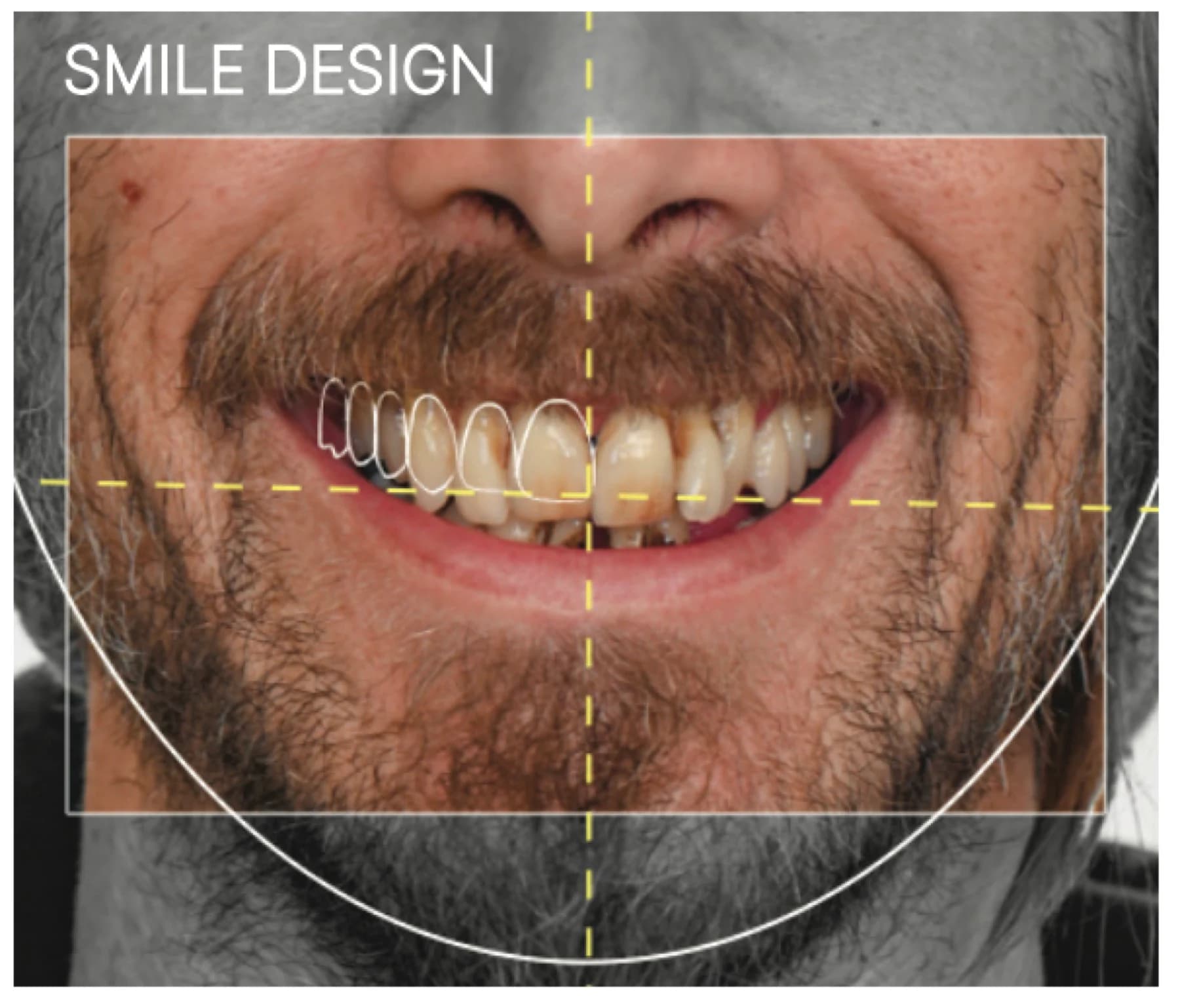
That is the reason the tooth is designed before the implant. The shape and length of the new front tooth are planned on a photograph of your smile with smile design, the implant is positioned in the 3D scan to support that tooth, and the surgery is carried out with X-Guide or Navident EVO dynamic navigation, which tracks the drill against the plan in real time. Our review of guided, navigated and robotic surgery explains what those systems can and cannot promise.
The gum between the teeth, and why two front implants side by side are harder
The papilla — the point of gum that fills the space between two teeth — is supported by bone. A classic 1992 study of 288 sites between natural teeth found that when the distance from the contact point of the teeth to the bone was 5 mm or less, the papilla was present almost every time; at 6 mm it was present 56% of the time, and at 7 mm or more, 27% of the time or less. Beside a single implant, the bone attached to the neighboring natural tooth still helps support the papilla on that side.
Between two implants the picture changes. A 2003 study of 136 papillae between adjacent implants found an average of only 3.4 mm of soft tissue above the bone, with a range of 1 to 7 mm, and its authors urged great caution when placing two implants next to each other in the smile. When two neighboring front teeth are missing, it is therefore often worth considering one implant supporting two crowns, or implants separated by a bridge tooth, rather than two implants side by side. The right answer depends on the bone and the bite, and it is decided in the plan, not after the gum has flattened.
The crown, the abutment and the color of the gum
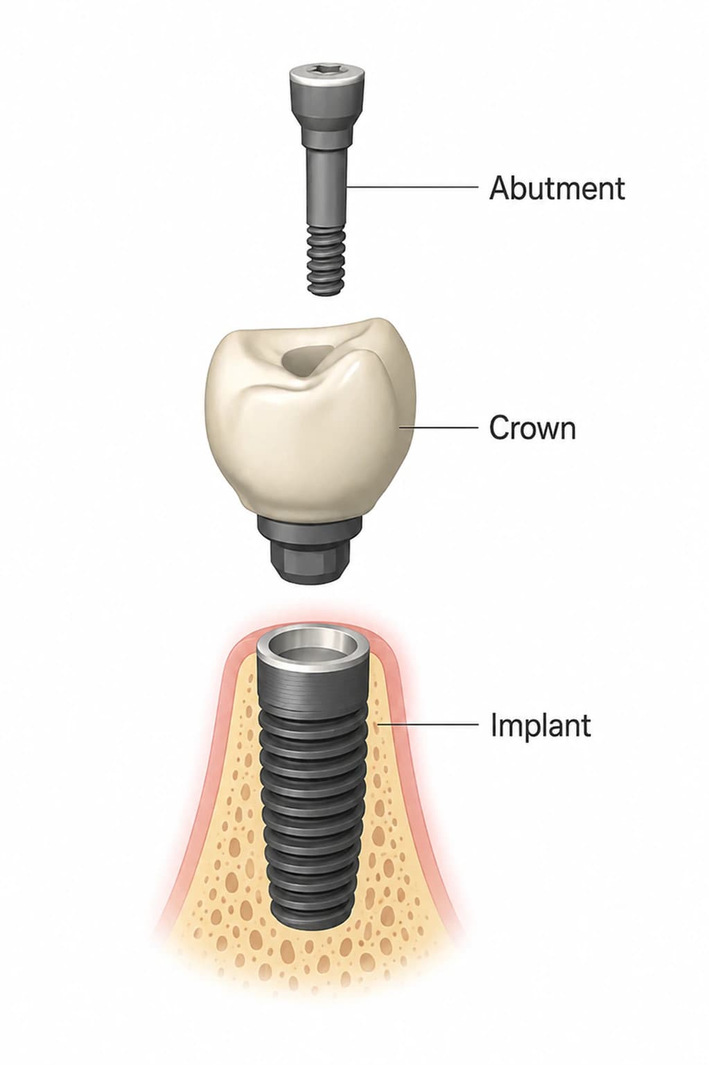
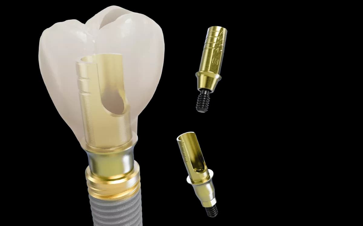
Healthy, thick gum hides a titanium implant completely. Thin gum can let a gray shadow show through, which is where a zirconia abutment, or in some mouths a zirconia implant, is worth discussing. Our article on gum grafting around implants goes through the color studies, and why adding gum thickness is sometimes part of a front-tooth plan.
The crown is where a front tooth is won or lost at a conversational distance. Your zirconia crown is designed, shaded and finished in our own lab a few steps from the treatment chair, so the lab team can compare it with your neighboring teeth in person rather than from a photograph. Where it can be, the crown is screw-retained, which lets it be removed for cleaning, adjustment or replacement without cutting it off, and leaves no cement under the gum. Before the final crown, a temporary crown shaped to guide the gum often comes first; the article on temporary teeth while an implant heals explains why that stage improves the final result.
The teeth around an implant keep moving, even in adults
An implant fused to bone stays exactly where it was placed. The natural teeth beside it do not: they continue to erupt and drift slowly throughout life. In a study of 28 patients with front-tooth implants, every patient in the older group, aged 40 to 55, developed a vertical step between the implant crown and the neighboring teeth, ranging from 0.12 to 1.86 mm over an average of 4.2 years — to the same extent as young adults whose growth was nearly complete.
Most of those steps are small. In a prospective study of 39 front implant crowns followed for up to six years, 51% showed no step at all, 36% a slight one under 0.5 mm, and only one crown more than 1 mm. A 2025 study following 133 front implant crowns for 11 to 21 years found some step in 68% of them, but only 8% of 1 mm or more; patients with a step of 0.5 mm or more were more likely to notice it, and most patients were unaware of any difference. The same study found that color mismatch between the crown and the natural teeth increased over the years. Implant survival was 99% and crown survival 92%.
The practical message is that a front implant crown may need to be remade one day because the teeth around it have changed, not because anything failed. A screw-retained crown makes that a lab visit rather than a surgical problem. For teenagers, whose jaws are still growing, the change is larger, and the implant is generally deferred until growth has finished, with the space held in the meantime.
Implant or bridge for a single front tooth
Our implants versus bridges page covers the general comparison. For a single missing front tooth there is one alternative worth knowing about specifically: a bonded bridge with a single wing glued to the back of one neighboring tooth, which needs little or no drilling of healthy teeth. Long-term case reports of all-ceramic versions describe incisor replacements that did not come loose and kept the gum under the false tooth healthy, in one case followed for 26 years, and their authors consider them a standard of care for a single missing incisor, often making an implant unnecessary for that indication.
A bonded bridge suits some mouths better than an implant: a young patient still growing, a site with very little bone, or a bite that leaves room behind the front teeth. It suits others less well, for example a deep bite, heavy grinding, or neighboring teeth that already need crowns. When a bonded bridge is the better fit, saying so is part of an honest consultation, and your general dentist can provide it.
What moves the cost of a front tooth implant
The parts of a front tooth implant are the same as for a molar, but the work around them often is not. A front tooth is more likely to need a graft to rebuild the thin front wall, sometimes a soft-tissue graft to thicken the gum, a temporary crown stage to shape the gum, a custom abutment, and more time in the lab matching shade and translucency. Each of those is a separate line on an honest quote. Our single tooth implant cost guide explains the components, and the Fremont cost page explains why a front tooth is not automatically the expensive one.
We do not publish a fee for single-tooth implants, because the number depends on what your scan shows. The consultation and CBCT scan are free, the plan is photographed and measured before anything is agreed, and the quote is itemized in writing. If you are weighing a front-tooth plan from another office, you are welcome to bring it for a second opinion.
Written and medically reviewed by Dr. Sam Jain, DMD, MS, and Dr. Arpana Gupta, DDS, MDS, who own this practice. Reviewed
This article is patient education, reviewed by Dr. Sam Jain, DMD, MS, and Dr. Arpana Gupta, DDS, MDS, and is not a substitute for an exam. Treatment recommendations are made only after an in-person consultation and 3D imaging.