Gum Grafting Around Dental Implants: Receding Gums, Metal Showing, and When Soft-Tissue Grafts Help
The gum around an implant does two jobs: it seals the implant from the mouth, and it hides the metal. When it is thin, missing its firm band of tissue, or pulls back, both jobs suffer. Soft-tissue grafting can often restore them — here is what causes the problem, what the evidence says grafts achieve, and where their limits are.

Written and medically reviewed by Dr. Sam Jain, DMD, MS, and Dr. Arpana Gupta, DDS, MDS
Published
In this article
- 1Why the gum around an implant matters
- 2Firm, keratinized gum: how much difference does it make?
- 3Thin gum, gray show-through and bone loss
- 4Why gums recede and metal starts to show
- 5What a gum graft around an implant involves
- 6What coverage can realistically achieve
- 7Changing the restoration instead of, or as well as, the gum
- 8Who is a good candidate
- 9How we approach receding gums around an implant
Why the gum around an implant matters
Around a natural tooth, the gum is attached to the root by fibers. Around an implant, the soft tissue forms a seal against the abutment instead, and that seal depends on the quality and quantity of the tissue. Two measurements come up in almost every conversation about it. One is the width of keratinized mucosa — the firm, pale pink band of gum that resists brushing and movement, as opposed to the softer, redder tissue further from the teeth. The other is mucosal thickness: how much tissue covers the implant and abutment from the outside.
Neither is a simple pass-or-fail measurement, and the evidence is more nuanced than some sales pitches suggest. But both influence how easy the implant is to clean, how stable the gum line stays, and whether any gray metal shows through. Those are the reasons a graft is sometimes recommended.
Firm, keratinized gum: how much difference does it make?
The research on keratinized tissue points in a consistent direction with honest caveats. A 2025 meta-analysis of 30 studies found that implants with at least 2 mm of keratinized mucosa had less plaque, less gum inflammation, less bleeding and slightly less bone loss than implants with less. A four-year study found that patients with a narrow band had more plaque, more bleeding and more brushing discomfort, and a 20-year study of implants in the lower back jaw found that those surrounded by mobile tissue rather than firm gum had more bone loss, bleeding, recession and peri-implant disease; half of those patients needed additional treatment, compared with 5% of patients with firm tissue.
The caveat is that the evidence is not uniform. A 2022 meta-analysis that applied stricter criteria rated the evidence for a narrow band as a risk factor for peri-implant disease as low, with only plaque levels clearly different. And a 25-year study found that the width of firm tissue was associated with long-term stability, but not with long-term brushing discomfort. The fair summary is that firm tissue helps, particularly for patients who find an area hard or uncomfortable to clean, but a narrow band on its own is not a reason for surgery.
Thin gum, gray show-through and bone loss
Mucosal thickness matters for appearance first. In a laboratory study, titanium produced the most visible color change in the tissue covering it; zirconia caused no visible change once the tissue was 2 mm thick, and at 3 mm the eye could not distinguish any of the materials tested. In a randomized clinical trial, all-ceramic restorations on ceramic abutments produced less visible gum discoloration than metal-ceramic crowns on titanium or gold abutments. A consensus workshop in 2022 concluded that thick mucosa tended to give better esthetic results and that the connective tissue graft remains the standard way to increase thickness.
Thickness also appears to matter for bone. In a one-year trial, implants surrounded by gum 2 mm thick or less lost up to about 1.5 mm of crestal bone despite being placed slightly above the bone, while implants with thick tissue lost very little. A 2018 systematic review found that grafting to increase mucosal thickness with the patient's own tissue was associated with significantly less marginal bone loss, and grafting to gain firm tissue with better bleeding scores and higher bone levels.
Why gums recede and metal starts to show
Recession around implants — a gum line that creeps up and exposes the abutment or even the implant's collar — is more common than most people realize. In a 2022 study of 176 single implants in the smile zone, some degree of this soft-tissue dehiscence was present at 56.8% of implants. It was associated with an adjacent implant, longer time in function, thin tissue, a narrow band of firm gum and a greater distance from the implant to the outer bone. After immediate implants in the front of the upper jaw, a systematic review found recession of more than 1 mm at a median of 26% of sites.
The single most important cause, according to a 2022 international consensus workshop, is an implant placed too far toward the lip or cheek; thin tissue came second. That is the reason prevention matters more than repair. Implants we place are planned in 3D and placed with guided navigation so the position leaves room for bone and gum on the outside, and when the tissue is thin, a connective tissue graft at the time of an immediate implant, which the same workshop identified as protective, is one of the options we consider.
Other contributors include bone loss from peri-implantitis, trauma from aggressive brushing, and crowns whose contours push the gum away. Recession that comes with bleeding, pus or a deepening pocket is an infection until proven otherwise, and our article on peri-implantitis treatment covers that path.
What a gum graft around an implant involves
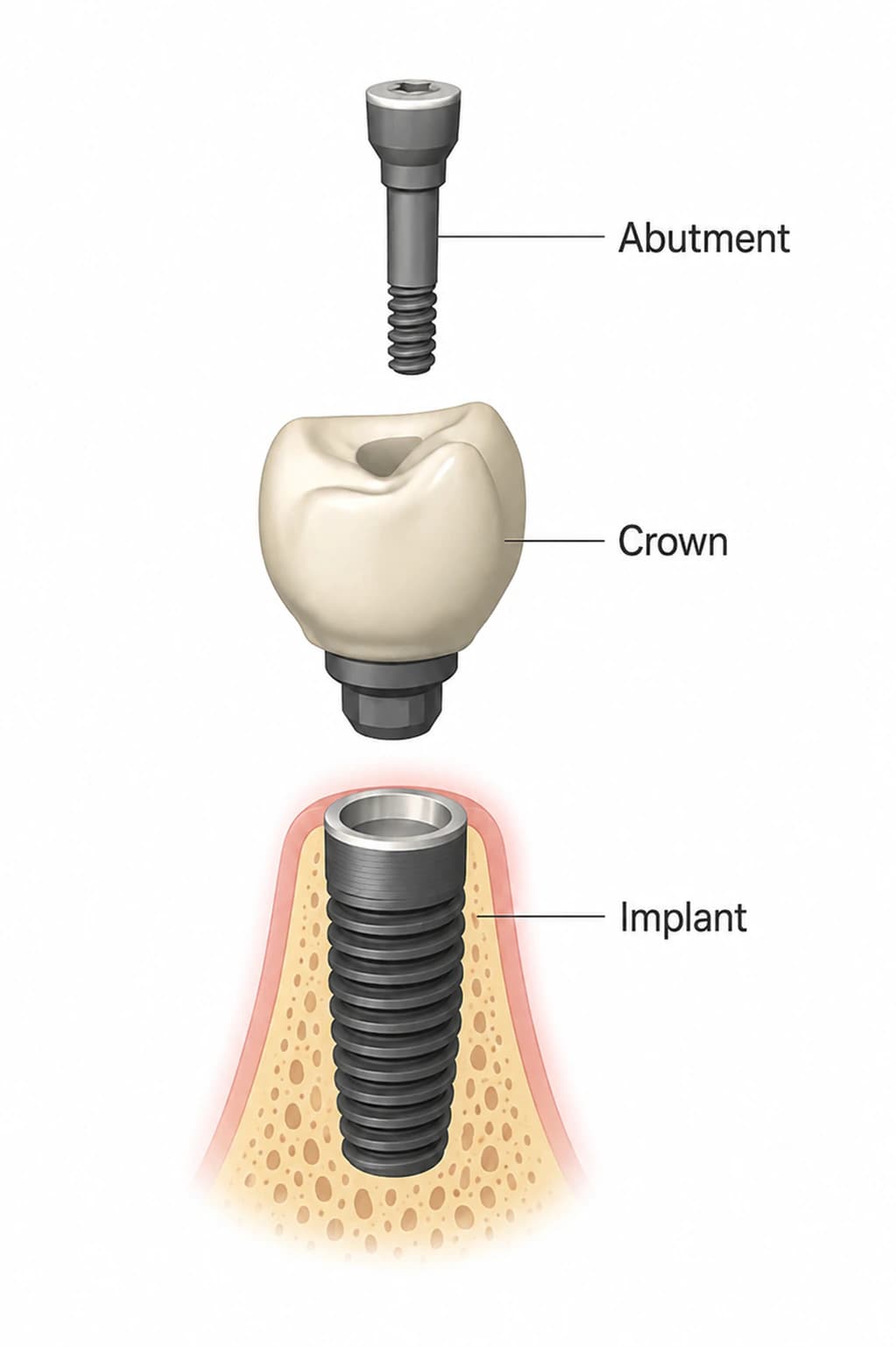
There are two broad aims, and the technique follows the aim. To add a band of firm gum, a free gingival graft — a thin strip of tissue taken from the palate — is placed at the implant; a systematic review found it gained about 0.9 mm more firm tissue than soft-tissue substitutes, although substitutes of animal origin came close and reduced surgical time and discomfort. To add thickness or cover a recession, a connective tissue graft is placed beneath the existing gum, often combined with moving the gum toward the crown, and sometimes with the crown or abutment removed or reshaped so the tissue has room.
The procedure is done under local anesthetic, usually in one visit, and the palate donor site is typically the sorer of the two areas for the first several days. Where it is appropriate, a soft-tissue substitute avoids the donor site altogether. The practice's gum graft consent explains the specific risks, including partial loss of the graft, incomplete coverage and the possible need for further grafting.
What coverage can realistically achieve
Covering recession around an implant is harder and less predictable than covering recession on a natural tooth, and the published evidence comes from a small number of studies. In a prospective study of shallow recessions at single upper implants treated with a connective tissue graft, complete coverage was maintained in 8 of 13 cases at five years, 62%, with an average of 86% of the recession covered and high esthetic ratings from patients. A review of coverage stability suggested waiting at least six months after treatment before judging the final gum position, because the margin settles over that period, and found tissue thickness to be the best predictor of a stable result.
Coverage is less achievable when the implant itself sits too far outward, when bone is missing on the outer surface, or when the recession is deep. In those situations the honest options may include accepting a small change, remaking the crown to disguise it, adding bone as well as soft tissue, or, when appearance matters most and the implant cannot be improved, replacing the implant in a better position. Our article on implant removal and replacement describes that last route.
Changing the restoration instead of, or as well as, the gum
Sometimes the most effective step is on the tooth side. A crown that bulges at the gum line can be reshaped so it supports the tissue rather than pushing it away. A titanium abutment that shows through thin gum can be replaced with a zirconia one, which the color studies suggest is far less visible through tissue 2 mm or more thick. A crown can be made with pink porcelain at the neck to mask a longer tooth where coverage is not realistic. Because our lab designs abutments and crowns in the building, those changes can be planned together with any grafting.
Who is a good candidate
Grafting tends to help most in a few situations: an implant in the smile zone where metal or a gray shadow is visible, a shallow recession around an otherwise healthy and well-positioned implant, an area that bleeds or hurts when brushed because it lacks firm tissue, and thin tissue at the time an implant is placed. It helps least when the underlying problem is an implant placed too far outward, active peri-implantitis that has not been treated, or a patient who smokes heavily.
Because the evidence varies by situation, the decision should be made on the specific implant, with photographs, measurements and a 3D scan showing the bone on the outer side. A narrow band of firm tissue that causes no symptoms may simply be watched.
How we approach receding gums around an implant
At a free consultation, we examine the gum around the implant, measure the band of firm tissue and the recession, check for inflammation and look at the implant's position and the bone on a 3D CBCT scan. That tells us whether the problem is the tissue, the restoration, the implant's position, or an infection, and each leads to a different recommendation. You receive the options and their realistic outcomes in writing, including when a graft is unlikely to give you the result you want.
Dr. Sam Jain and Dr. Arpana Gupta perform grafting procedures in the office, and any change to the abutment or crown is designed in our own lab. Whether the implant was placed here or elsewhere, you can book a consultation here.
Written and medically reviewed by Dr. Sam Jain, DMD, MS, and Dr. Arpana Gupta, DDS, MDS, who own this practice. Reviewed
This article is patient education, reviewed by Dr. Sam Jain, DMD, MS, and Dr. Arpana Gupta, DDS, MDS, and is not a substitute for an exam. Treatment recommendations are made only after an in-person consultation and 3D imaging.