Clinical Case: Single Implant With GBR at a Maxillary Lateral Incisor
A walk-through of one of the trickiest single-tooth situations in dentistry — a front tooth in thin bone — and how guided bone regeneration made a natural, stable result possible.

Written and medically reviewed by Dr. Sam Jain, DMD, MS, and Dr. Arpana Gupta, DDS, MDS
Published · Updated
The Short Answer: Yes, a Thin Front-Tooth Site Can Still Get One Implant
The reader question behind this case is usually some version of: my dentist says there isn't enough bone for an implant at my upper front tooth — do I really need a graft, and will it still look natural? For a maxillary lateral incisor, the honest answer for many patients is yes to the graft and yes to the natural result, and guided bone regeneration (GBR) is what bridges the two. GBR rebuilds the missing bone at the same time the implant goes in, so one thin site becomes a stable one.
The lateral incisor is a small tooth in a demanding spot. It sits at the corner of your smile, the bone over its root is often paper-thin to begin with, and once a tooth is lost that outer wall tends to collapse inward. Place an implant into that thinned ridge without rebuilding it and you risk exposed metal threads, a gray shadow through the gum, or a tooth that looks longer than its neighbor. GBR is the step that prevents all three.
This is one real case, shared as patient education. Every mouth is different, so your own plan comes from an exam and a 3D scan — but the principles here are the same ones we use at the Center for Implant Dentistry on aesthetic-zone single implants every week.
The Starting Point: A Narrow Ridge at the Corner of the Smile
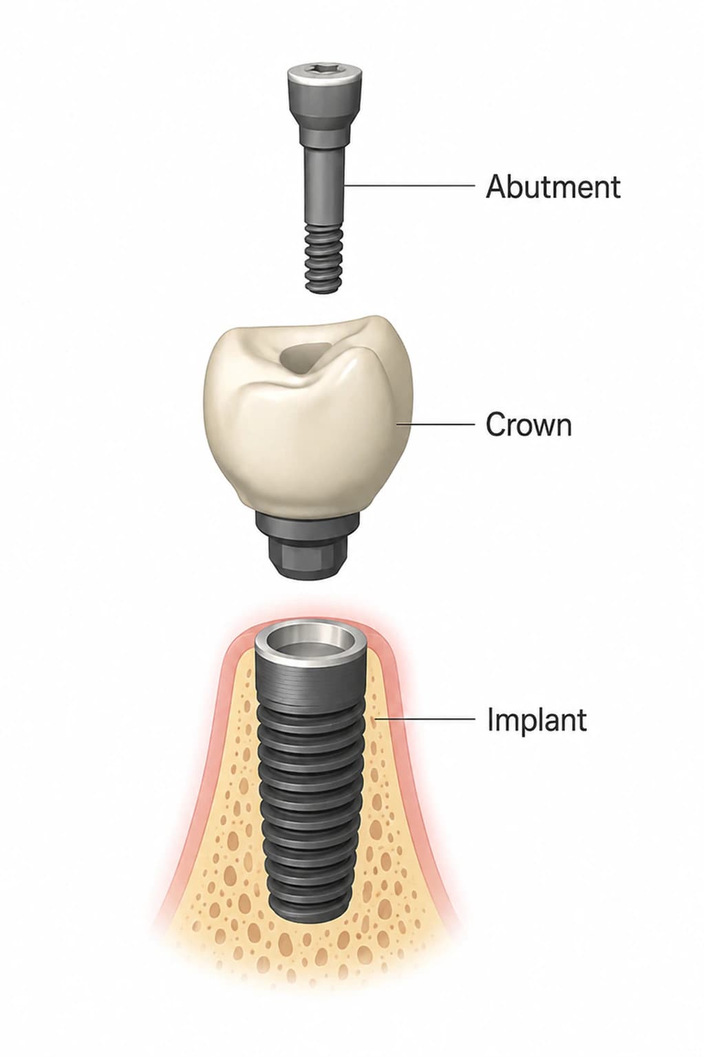
The patient came in having lost an upper lateral incisor. On the free 3D CBCT scan, the story was clear before anyone picked up an instrument: the ridge was there in height but pinched in from front to back, with a thin, partly resorbed outer (buccal) plate of bone. That's the classic lateral-incisor picture. It's exactly the anatomy that makes this tooth harder to restore than the bigger, blockier teeth around it.
Planning happened on the scan, not on the day. Dr. Sam Jain — who holds a DMD plus a master's in mechanical engineering, and is an ICOI Master — mapped where the implant had to sit to support a natural-looking tooth, then measured how much bone was missing on the outer face to get it there. Because the same team plans, places, and restores every case here, the implant position was chosen for the final crown from the very first step, not reverse-engineered later. That planning is done with guided-surgery systems (X-Guide and Navident EVO) so the drill follows the plan, which matters more at a front tooth than almost anywhere else. You can see the broader single- and few-tooth workflow on our single and multiple implants page.
The Procedure: One Appointment, Implant and Graft Together
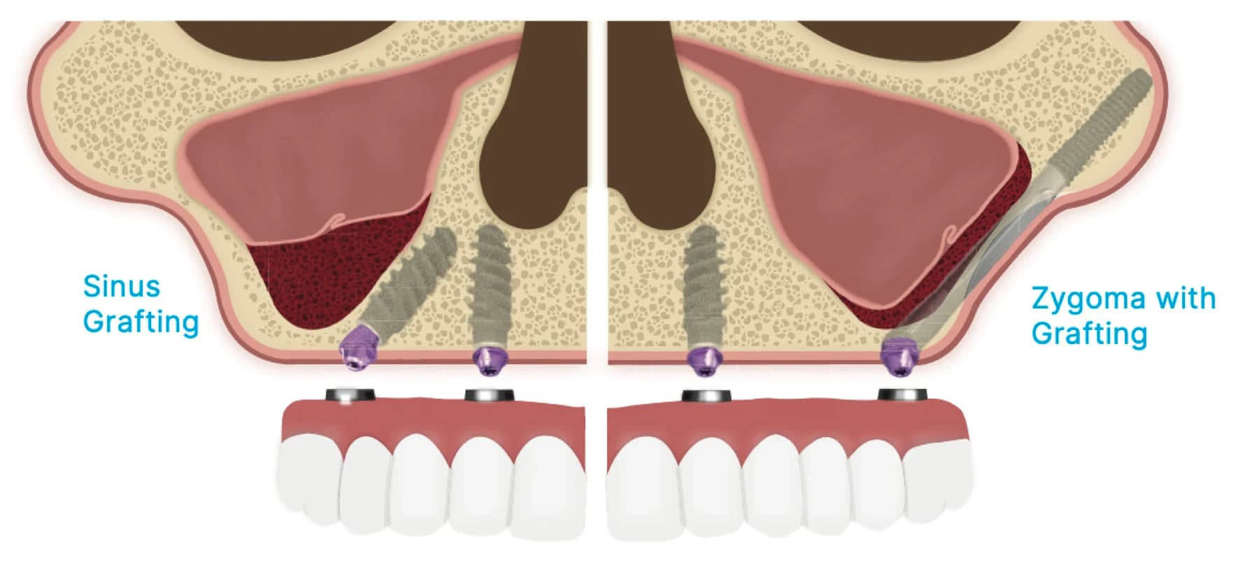
This case used a simultaneous approach — the implant and the bone graft in the same visit — which is the standard when there's enough native bone to hold the implant steady while the graft heals around it. The tooth site was opened gently, the implant was placed into solid bone at the planned position, and the thin outer wall was rebuilt over the exposed threads. If the ridge had been too deficient to anchor an implant at all, we'd have staged it: graft first with our bone grafting protocol, let it mature, then place the implant later. Your surgeon confirms which path your bone allows.
The GBR itself is a layered repair. Graft particles (a bone substitute, often mixed with the patient's own bone) are packed against the defect to give new bone a scaffold to grow into. A collagen barrier membrane is laid over the top like a tarp — its job is to hold the graft in place and keep faster-growing gum tissue from invading the space while slower bone catches up. The gum is then closed over it to heal. It's a quiet, methodical step, done here under IV sedation so the patient is comfortable and remembers little of it.
A note on membrane choice, because it's a real clinical decision: resorbable collagen membranes dissolve on their own, so there's no second surgery to remove them, and in comparative studies they expose or get infected far less often than the older non-resorbable type — on the order of about 5% versus roughly 20%. For most single anterior sites, that lower complication profile is why a resorbable membrane is the workhorse.
Why the Result Looked Natural — and What the Evidence Says
Rebuilding the outer bone wall does more than protect the implant; it's what gives the gum something to drape over. Bone is the foundation the gumline sits on, so restoring buccal thickness is how you avoid the tell-tale gray show-through and the receded, too-long look that gives a front-tooth implant away. Once the implant fused, it was restored with a custom crown milled on our in-house CAD/CAM zirconia system — final custom teeth in roughly a day for single-tooth cases, rather than months in a temporary. The finished lateral incisor matched its neighbor in length, contour, and how the light caught it.
The research backs this up as a predictable procedure, not a gamble. Across studies of GBR used to correct bone defects around implants, implant survival is high — commonly reported around 95% and up (one systematic review put the pooled figure near 95.7%), and single-implant survival in general runs about 95% or better over the long term. Those are population averages, and your own odds depend on your bone, your bite, your health, and your hygiene, which your doctor will go over with you.
The reason we treat the lateral incisor with this much care is that the margin for error is small and the payoff for getting it right is a tooth nobody can pick out of a smile. If you were told a front-tooth site is off the table, it's worth a second look — the same skills scale up to full-arch cases and the hardest rescue and complication work we do. The free consultation and 3D scan are the honest way to find out what your own site needs.
Written and medically reviewed by Dr. Sam Jain, DMD, MS, and Dr. Arpana Gupta, DDS, MDS, who own this practice.
This article is patient education, reviewed by Dr. Sam Jain, DMD, MS, and Dr. Arpana Gupta, DDS, MDS, and is not a substitute for an exam. Treatment recommendations are made only after an in-person consultation and 3D imaging.