Osseointegration: How Bone Actually Bonds to a Titanium Implant
Bone does not grow onto an implant the way glue sets. It first resorbs the very bone that holds the implant, then rebuilds. Understanding that sequence explains almost every rule I follow.

Written and medically reviewed by Dr. Sam Jain, DMD, MS, and Dr. Arpana Gupta, DDS, MDS
Published
A definition that has changed since 1977
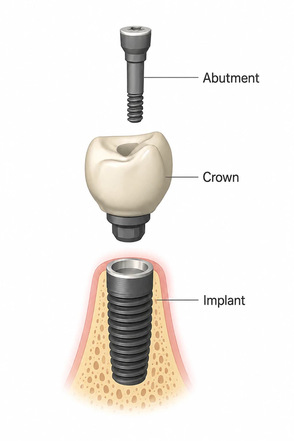
The word was coined by Per-Ingvar Brånemark's group in Gothenburg, whose ten-year report on osseointegrated implants in the toothless jaw appeared in 1977, and it was defined operationally in the 1981 paper by Adell and colleagues: a firm, direct and lasting connection between living bone and a screw-shaped titanium fixture, with no soft tissue between them. Three conditions were attached — gentle surgical technique, a long healing time, and a proper distribution of stress once in function. Forty-five years of research have refined every clause of that sentence without overturning any of it.
In 2001 Albrektsson and Johansson separated three words that are still confused in patient conversations. Osteoinduction is the recruitment of immature cells and their stimulation into bone-forming cells; it drives most fracture healing. Osteoconduction is bone growing along a surface, which is what a titanium implant permits and what copper, silver or bone cement do not. Osseointegration is the stable anchorage that results from direct bone-to-implant contact. In the jaws, it is the only mode of anchorage with a long record of high success rates; whether it matters as much for a hip replacement is, they noted, still debated.
The most interesting revision came from Albrektsson himself in 2019, writing with Wennerberg. Their narrative review argues that an oral implant is, biologically, a foreign body, and that osseointegration is best understood as the tissue's protective response to it: given adequate stability, bone forms around titanium to shield the body from it. That reframing matters because it predicts what we see clinically — that the bond is dynamic, that it is maintained by ongoing remodeling, and that it can be lost if the balance between the implant and the tissue around it is disturbed. It also explains why the implant surface has become the most studied variable in the field.
The first six weeks, step by step
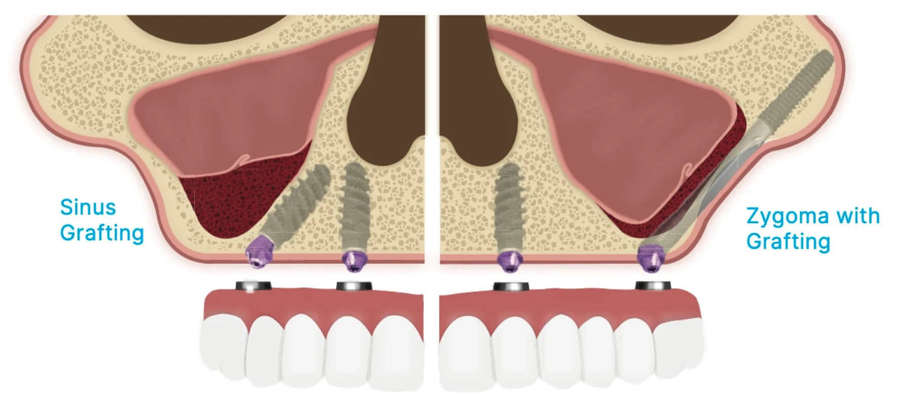
The clearest description of the cascade is Davies' 2003 paper, which divides healing into three phases. First, osteoconduction: bone-forming cells migrate to the implant surface through the remains of the blood clot, a process driven by platelet activation at the surface itself. Second, de novo bone formation: those cells lay down a mineralized matrix directly on the implant, equivalent to the cement line in natural bone, which is what produces contact osteogenesis and, on a suitable surface, a true bone bond. Third, remodeling, which is slower and continues for the life of the implant. Davies' central point is that the most important steps in the first two phases are profoundly influenced by the microscopic texture of the implant surface.
Berglundh, Abrahamsson, Lang and Lindhe watched this happen in 2003 using an implant with a deliberately hollowed thread chamber, placed in twenty Labrador dogs and examined from two hours to twelve weeks. The empty chamber filled first with clot, then with granulation tissue, then with a provisional matrix. Bone formation began during the first week, and — this is the finding I explain to every patient who asks why they must be careful in the early weeks — between weeks one and two, the bone immediately against the thread pitch, the bone that had provided the implant's initial mechanical grip, was resorbed and replaced with newly formed bone. The implants stayed clinically stable throughout, but for a short period their stability was borrowed from new bone that was still being built.
The human timeline was measured directly by Lang and colleagues in 2011. Small titanium devices were placed behind the last molars of 49 volunteers and retrieved with a core of surrounding tissue after 7, 14, 28 or 42 days. New bone formation started in the first week in the trabecular regions and increased steadily. The proportion of the surface in direct contact with new bone was 12.2% at two weeks and 32.4% at four weeks on a standard sandblasted, acid-etched surface, and 14.8% and 48.3% on the chemically modified hydrophilic version of the same surface. By 42 days the two had converged at 61.6% and 61.5%. Six weeks is therefore a real biological milestone rather than a convention, and the hydrophilic surface's advantage is a matter of speed in the first month rather than of the final result.
The stability dip, and the number that governs loading
Meredith's 1998 paper gave the field the vocabulary it still uses. Primary stability is mechanical: it comes from the fit between the implant and the bone at the moment of placement, and depends on bone quality, bone quantity, implant design and surgical technique. Secondary stability is biological: it is the stability added by bone formation and remodeling at the interface. Between the two lies the dip that Berglundh's dogs demonstrated, when the mechanical grip is being resorbed faster than the biological bond is being built. Meredith also described resonance frequency analysis — the small transducer that vibrates the implant and reports an implant stability quotient — and Sennerby and Meredith's 2008 review in Periodontology 2000 established it as the standard non-destructive way to follow that curve.
Whether an implant can carry load during the dip depends on how much it moves. Szmukler-Moncler and colleagues reviewed the experimental literature in 1998 and reached a conclusion that overturned the original three-to-six-month rule: early loading in itself was not found to be harmful to osseointegration. Only excessive micromotion at the interface was directly implicated in fibrous encapsulation — the scar-tissue sleeve that is the histological definition of failure. The tolerated threshold was not zero. It lay somewhere between 50 and 150 microns. Below it, bone forms; above it, scar forms; and the whole art of immediate loading is keeping every implant on the right side of that line while it heals.
The clinical proxy for that threshold is insertion torque. Ottoni's randomized study of 46 single implants in 2005 restored one implant in each patient with a provisional crown within 24 hours and left the other to heal conventionally. Ten of the immediately restored implants failed, and nine of those ten had been placed with the minimum insertion torque of 20 Ncm. The relative risk of failure fell by about 20% for every additional 9.8 Ncm of torque. That is why insertion torque is measured on every implant I place, and why a reading below my threshold turns a same-day tooth into a two-stage plan on the spot. The provisional is the easy part; the number decides whether it is safe.
Why the surface matters — and where the claims outrun the data
Wennerberg and Albrektsson's 2009 systematic review is the reference point for surface texture. Of 1,184 publications identified, 1,064 had to be discarded because they did not actually measure the bone response to a characterized surface, and 20 more dealt mainly with calcium-phosphate-coated or zirconia implants, leaving 100 papers. Those showed clearly that surface topography influences bone response at the micrometer scale, with moderately rough surfaces — an average roughness of roughly one to two microns — producing stronger bone responses than smoother or rougher ones in a number of studies. They also found that most papers characterized their surfaces inadequately, that a surface called rough in one study was called smooth in another, and that measurement methods needed standardizing. Buser's 2017 history of the field credits the shift to moderately rough surfaces as the change that made early and immediate loading protocols possible at all.
Beyond microroughness the evidence thins quickly, and it is worth being specific about where. Albrektsson and Wennerberg's 2019 review concludes that an isotropic, moderately rough surface has shown better clinical results than the minimally rough or very rough surfaces used previously, but that there is no clinical evidence supporting any particular nanoscale pattern, that a benefit from surface chemistry is possible but unproven, and that hydrophilic surfaces have encouraging animal data without clinical proof of better outcomes. Rupp's 2018 review reaches a similar place: hydrophilic surfaces demonstrably improve early blood contact and wound healing, but a surface engineered for one tissue interface with predictable selectivity is, in their words, still far away. Smeets and colleagues' 2016 survey of surface modifications is a fair catalog of what is being tried — laser ablation, crystalline deposition, protein and growth-factor coatings — and an honest account of how much of it remains experimental.
So when a manufacturer's brochure describes a surface as bioactive, or nanostructured, or accelerating integration, I read it against those two reviews. The moderately rough textured surface is backed by decades of clinical outcomes. The refinements on top of it may be real, and Lang's human data suggest the hydrophilic version does buy speed in the first month, but nobody has yet shown that a patient loses fewer implants because of them. I choose implant systems on the documented part and treat the rest as a possible bonus. The materials page explains which systems we use and why.
What this means for your timeline
Putting the biology together gives a sequence rather than a single healing time. Days one to seven: clot, then a provisional matrix, with the first new bone already appearing in spongy bone. Weeks one to two: the borrowed mechanical grip begins to be resorbed and replaced, which is the window in which movement matters most. Weeks four to six: bone-to-implant contact climbs steeply, roughly doubling in Lang's volunteers between day 14 and day 42. Beyond six weeks: remodeling continues for years, and the interface is maintained by the same turnover that maintains every other bone in the body.
The protocol I choose for a given implant follows that curve. An implant with high insertion torque, in dense bone, splinted rigidly to others across the arch, can carry a provisional from the first day because its micromotion stays under the threshold — that is the biology behind same-day teeth, and it is why the decision is made at surgery with the torque wrench in hand rather than at the consultation. A single implant in soft upper-jaw bone, or one placed into a grafted site, is left unloaded through the dip and restored once secondary stability has been confirmed. Neither is the better protocol; they are the right protocol for different interfaces.
Your part in the first six weeks is mechanical, not medical. No chewing on a provisional that has been placed out of the bite, no probing the site with a tongue or a toothbrush that is too eager, and no smoking, which impairs the blood supply the whole cascade depends on. The what-to-eat guide turns that into a menu. If something feels loose in that window, it is a same-day phone call, because an implant moving during the dip is the one situation in which waiting makes the outcome worse.
Open questions I follow closely
The foreign-body framing of osseointegration is not universally accepted, and Buser's 2017 review lists it among the field's live controversies, alongside the debate about how peri-implantitis should be defined. If Albrektsson is right, then some marginal bone loss around implants is an immune equilibrium rather than an infection, which would change how we interpret a radiograph. That question is being worked out in the laboratory now and I would expect it to shape clinical guidance within the decade.
The practical gap is a better way to see secondary stability before it is tested by a bite. Resonance frequency analysis is useful but coarse. Work on peri-implant biomarkers, on ultrasound imaging of the interface, and on surface treatments that shorten the dip — including the ultraviolet photofunctionalization I discuss in the future-research review — is aimed at exactly that window. Until it matures, insertion torque and time remain the two instruments I trust, and the long-term survival data are the evidence that, used carefully, they are enough.
Written and medically reviewed by Dr. Sam Jain, DMD, MS, and Dr. Arpana Gupta, DDS, MDS, who own this practice. Reviewed
This article is patient education, reviewed by Dr. Sam Jain, DMD, MS, and Dr. Arpana Gupta, DDS, MDS, and is not a substitute for an exam. Treatment recommendations are made only after an in-person consultation and 3D imaging.