The Future of Dental Implants: Seven Lines of Research, Graded by How Close They Are to Your Mouth
Some of this is already in the operating room. Some of it is a decade away. The useful thing is to know which is which, and the literature is clearer about that than the marketing is.

Written and medically reviewed by Dr. Sam Jain, DMD, MS, and Dr. Arpana Gupta, DDS, MDS
Published
In this article
- 1How I grade a new technology
- 2Faster integration: ultraviolet light and growth factors
- 3When there is no bone: printed subperiosteal frames
- 4A different material: zirconia
- 5Precision: robots, planning software and photogrammetry
- 6Diagnosis before damage: biomarkers around the implant
- 7What I would put money on, and what I would not
How I grade a new technology
Every year brings a new implant surface, a new device and a new acronym, each described as a breakthrough. Most are not, and a few are, and telling them apart is a skill patients deserve to have explained rather than performed on them. I use a simple ladder. At the bottom is bench and animal work: a mechanism that works in a dish or a rat. Above it are human case series and retrospective cohorts: it has been done in patients and nobody has counted the failures properly yet. Above those are controlled trials, then systematic reviews of trials, and at the top are long-term outcome data — ten years, in this field — that show a patient actually kept more implants because of it.
Read that ladder against the survival-rate review and you will see the problem for any new technology. The standard treatment already has ten-year survival in the mid-nineties. To prove an improvement on that, a trial needs thousands of patients followed for a decade, and almost nothing new has that yet. So the honest grade for most of what follows is not “does it work” but “how much of the ladder has it climbed”, and I have tried to say exactly where each one stands. Where something is already used in our practice, I say that too, and I say what it is used for, because the indication is usually narrower than the brochure.
Faster integration: ultraviolet light and growth factors
Titanium ages. A sterile implant sitting in its packaging accumulates a film of hydrocarbons on its oxide surface that reduces its attraction for blood, proteins and cells. Aita and Ogawa's 2009 paper in Biomaterials showed that exposing titanium to ultraviolet light for up to 48 hours stripped that film: attachment, spread, proliferation and differentiation of bone cells rose by up to threefold, new bone formed on the treated implants in rats with almost no soft-tissue intervention, bone-to-implant contact approached 100% at four weeks, and the mechanical fixation of the implant was established four times faster. The effect tracked the removal of surface carbon rather than wettability as such, and they named it photofunctionalization. Funato, Yamada and Ogawa's 2013 retrospective series is the clinical follow-through: 168 photofunctionalized implants in 70 patients, treated for 15 minutes chairside just before placement, against 222 untreated implants in 95 patients. Healing time before loading was 3.2 months against 6.5, success was 97.6% against 96.3%, and the monthly gain in stability was markedly higher — despite the treated group using shorter and narrower implants. Chang's 2022 narrative review found the biology consistent and the clinical trials still too few and too focused on simple cases. Grade: convincing mechanism, one good retrospective series, no randomized trial with hard outcomes. It is a promising adjunct rather than a proven one.
Growth factors take the opposite route — changing the bone rather than the implant. Recombinant human bone morphogenetic protein-2, delivered on a collagen sponge, is the one with a real trial behind it: Fiorellini's 2005 multicentre randomized study placed it into 80 extraction sockets with at least half the facial bone missing, and the higher concentration produced significantly more bone than the sponge alone or no treatment, with a striking effect on new bone formation in the authors' words. It remains expensive and prone to post-operative swelling, which has kept it a tool for specific defects rather than a routine graft. Platelet-rich fibrin, the patient's own concentrated clot, is cheaper and widely used; Strauss's 2018 review found moderate evidence for benefit in ridge preservation and early osseointegration, and Miron's 2017 review found little randomized evidence that it adds bone in augmentation or sinus lifts. Grade for BMP-2: proven for its narrow indication. Grade for PRF: proven for soft tissue and socket healing, unproven for bone volume — which is exactly how I use it, and what the graft-materials review and our PRF consent form both say.
When there is no bone: printed subperiosteal frames
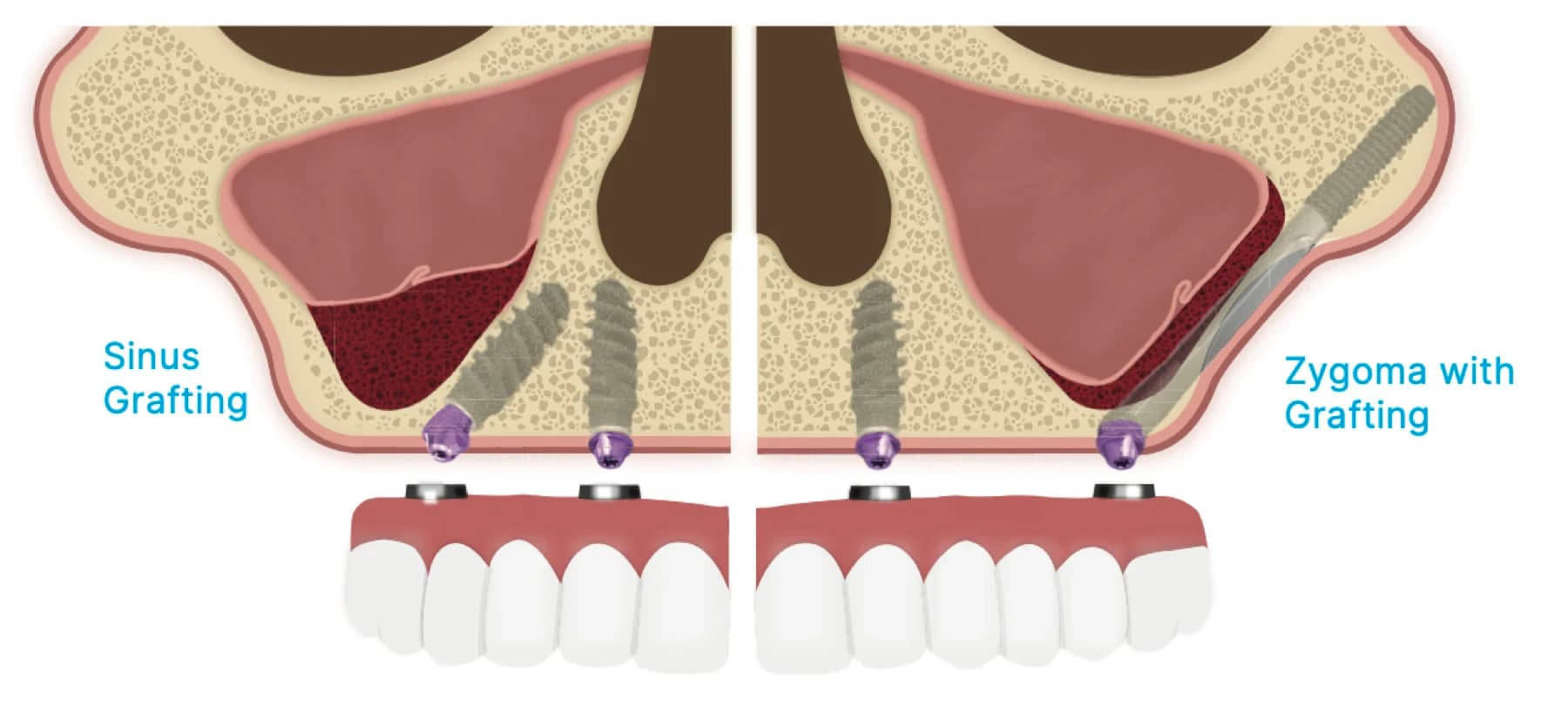
Subperiosteal implants — metal frames that sit on top of the jawbone under the gum rather than inside it — were tried in the 1960s and abandoned for good reasons that Buser's 2017 history records: questionable outcomes and no scientific documentation. They have returned in a form the original designers could not have imagined. Mommaerts' 2017 technical note describes the modern version: the patient's CBCT is segmented, a frame with integrated abutments is designed to the bone and to the planned teeth, and frame and teeth are printed in titanium alloy and polymer, so that a patient with extreme atrophy can be restored in one surgical session without any bone graft. Cerea and Dolcini's 2018 retrospective series followed 70 patients over 60 with laser-sintered custom frames for two years: survival was 95.8%, three frames were lost to recurrent infection, and prosthetic complications occurred in 8.9% of the survivors. Anitua's 2024 systematic review pooled thirteen studies and 227 implants with a mean follow-up of 21.4 months: 97.8% remained in function, but 25.6% showed partial exposure of the frame through the gum and 5.3% of patients had soft-tissue or persistent infection. Grade: a real option for the jaw with no bone, with two-year survival that is good and a soft-tissue complication rate that is not yet acceptable for a routine indication. It is a serious alternative to zygomatic implants in the most atrophic cases and needs five-year data before it is more than that.
A different material: zirconia
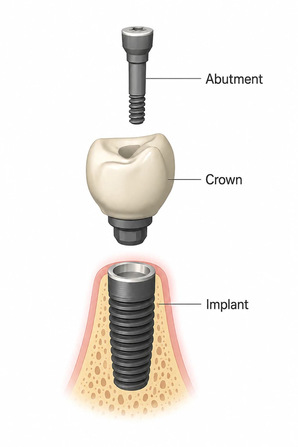
Ceramic implants are the second attempt at a metal-free implant, and this time the material — yttria-stabilized zirconia — is strong enough. Roehling's 2018 meta-analysis of eighteen clinical studies separated commercially available designs from experimental ones and found that the commercial one-piece implants had survival of 98.3% at one year and 97.2% at two years, a mean one-year bone loss of 0.7 mm, technical complications in 1.6%, implant fractures in 0.2% and biological complications in 4.2% — figures similar to titanium at the same time points. The 2018 ITI consensus put it carefully: one-piece zirconia implants can be recommended when appropriate clinical conditions exist, two-piece zirconia implants should be used with caution for lack of data, and zirconia bridges are less reliable than metal-ceramic ones. Grade: two-year data comparable to titanium, ten-year data absent, and a design constraint — the one-piece implant cannot be angled with an abutment after placement — that limits it to well-planned single sites. Our titanium-versus-zirconia page covers when a patient might choose it.
Precision: robots, planning software and photogrammetry
Robotic placement is the newest step in the accuracy story told in the guided-surgery review, and its grade is the same as there: Khaohoen's 2024 meta-analysis found the robotic subgroup reporting the smallest deviations of any approach, 0.81 mm at the entry and 1.71 degrees, from a handful of early series, with no long-term outcome data and no trial showing that the extra accuracy produces a longer-lived implant. Dynamic navigation, by contrast, has climbed most of the ladder, and is in daily use in our rooms.
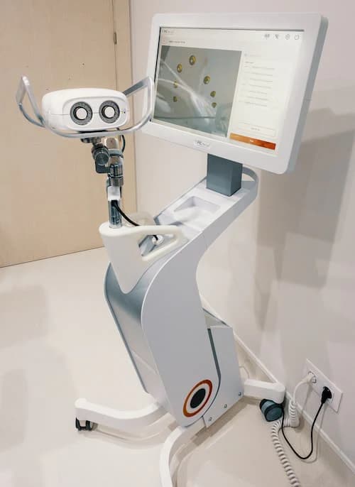
Photogrammetry — cameras that record the position of scan bodies on multiple implants in one capture — is the technology that would let a full-arch bridge be milled from a digital record with no physical impression. Its grade is a caution against assuming new means better. Revilla-León's 2021 laboratory study measured six implant positions on a model recorded by a conventional splinted impression, a photogrammetry system and two intraoral scanners against a coordinate-measuring machine: the conventional impression had the lowest three-dimensional discrepancy, the intraoral scanners were not significantly different from it in linear distortion, and the photogrammetry system tested had the highest discrepancy of all. A 2023 study from the same group, again measured against a coordinate-measuring machine, found a photogrammetry system within about two microns of the conventional method in trueness but with a precision spread nearly four times wider and an uneven discrepancy from one implant position to the next. The lesson stands. In our laboratory, a full-arch digital record is verified against a physical one before a final bridge is milled, and the 72-hour full-arch workflow is built on that verification rather than on trust in any single capture.
Diagnosis before damage: biomarkers around the implant
The one development I would most like to see mature is not a device but a test. Everything we use to diagnose peri-implantitis — the probe, the radiograph — measures destruction that has already happened. Alassy's 2019 review of biomarkers in the fluid that seeps around an implant describes what a real diagnostic would look like: levels of interleukin-1β, tumor necrosis factor-α and matrix metalloproteinase-8 that report current inflammatory activity rather than past bone loss. Chairside tests for MMP-8 already exist for periodontal disease. The review's honest conclusion is that these markers may have value in diagnosing peri-implant disease, that sampling methods and interpretation vary between studies, and that the research is insufficient to say whether any of them predicts progression. Grade: laboratory-validated, clinically immature, and the thing that would change practice most if it worked — because the peri-implantitis data show that prevention succeeds and treatment struggles, and a test that catches the disease before bone loss would move patients from the second group into the first.
What I would put money on, and what I would not
Photofunctionalization and hydrophilic surfaces will, I expect, quietly become standard — not because they will change ten-year survival, which is already high, but because they shorten the vulnerable window after surgery, and a shorter window is worth having. Printed subperiosteal frames will find a durable niche in the most atrophic jaws once the soft-tissue problem is engineered out. Biomarker tests will arrive, and when they do the recall visit will change from an inspection to a measurement. Navigation and robotics will converge, and the robot will win on tremor and lose on cost until the cost falls.
I would not bet on any of them retiring the fundamentals in the osseointegration review: a clean surgery, a stable implant, a respected healing period, a cleanable restoration and a patient who comes back. Every technology on this page is an attempt to make one of those five easier or more certain. None of them replaces one. When a new device is presented to you as doing so, that is the moment to ask where on the ladder it stands, and the answer is usually lower than the brochure.
Written and medically reviewed by Dr. Sam Jain, DMD, MS, and Dr. Arpana Gupta, DDS, MDS, who own this practice. Reviewed
This article is patient education, reviewed by Dr. Sam Jain, DMD, MS, and Dr. Arpana Gupta, DDS, MDS, and is not a substitute for an exam. Treatment recommendations are made only after an in-person consultation and 3D imaging.