Implant Troubleshooting by Dr. Sam Jain: Small Sinus Tract on a 3-Year-Old Implant
A little pimple on the gum next to an implant that healed years ago is a specific signal, not a random flare-up — here's how a surgeon reads it, and why the fix depends entirely on the cause.
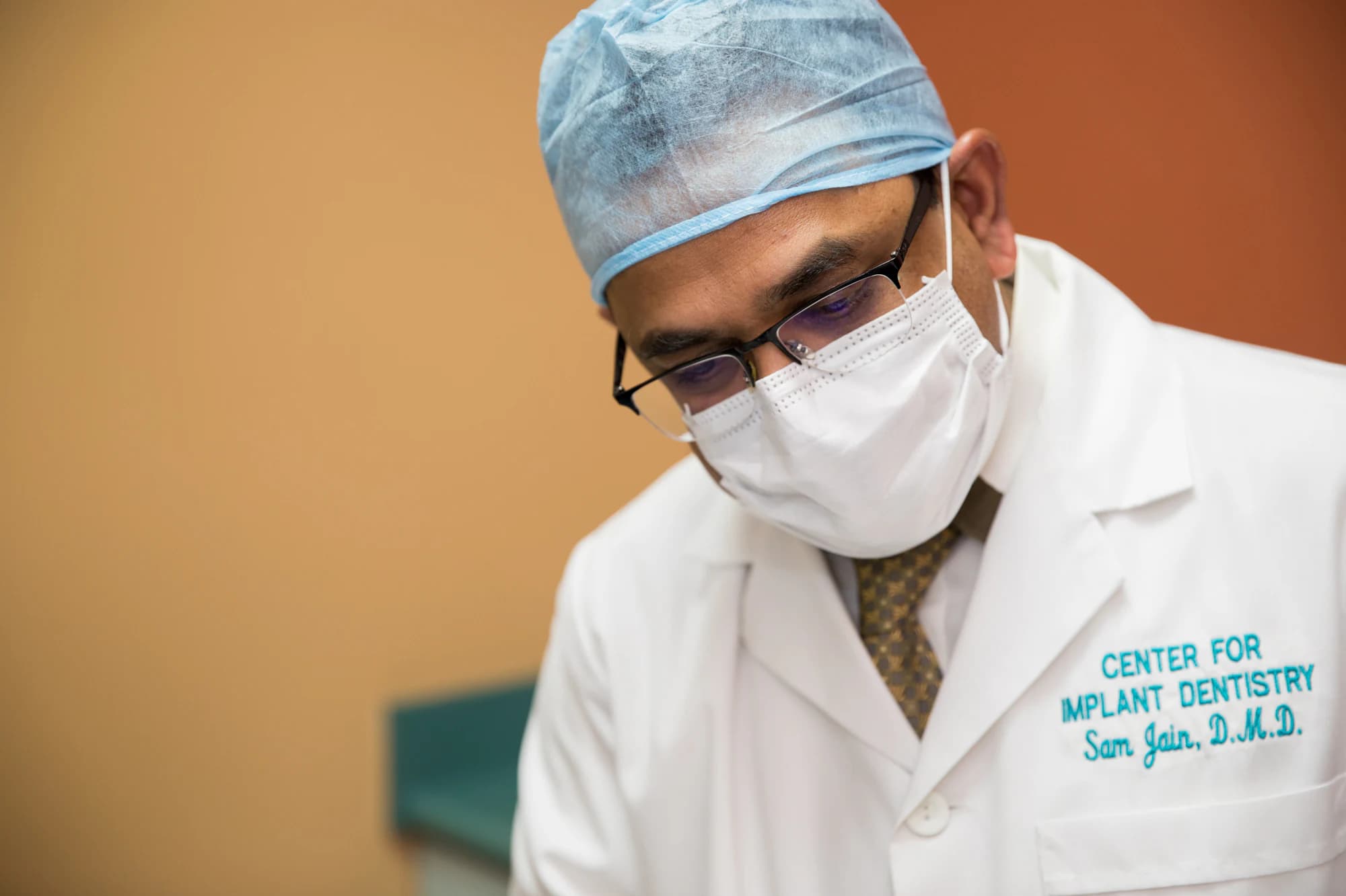

Written and medically reviewed by Dr. Sam Jain, DMD, MS, and Dr. Arpana Gupta, DDS, MDS
Published · Updated

What That Little Bump Actually Is
A sinus tract — patients usually call it a gum boil, a pimple, or a fistula — is your body's drainage channel. Somewhere below the surface there's a low-grade infection, and pus is finding the path of least resistance out to the gum. When it shows up next to an implant that healed cleanly and worked fine for three years, the tract itself isn't the problem. It's the messenger. The real question every time is: where is the infection coming from, and is the implant still solidly fused to bone?
Here's the part that reassures most people: a small, painless tract on a long-stable implant does not automatically mean you're losing the implant. Plenty of these trace back to a fixable source above or beside the implant rather than failure of the implant itself. But it is not something to watch and wait on, and it is not something a course of antibiotics alone will cure. Antibiotics can quiet the drainage for a week or two, then it comes right back — because the tablets never touched the actual source. You need someone to find the source, and that starts with a 3D scan.
So the honest triage is this: get it imaged, get it traced, and treat the cause. A tract that has been there for years, on an implant that's still rock-solid, usually has a good outcome. A tract that comes with a loose implant is a different, more urgent conversation. The scan tells you which one you're dealing with.
Why It Shows Up at Year Three, Not Year One
The timing throws people, because the implant integrated, the crown went on, and everything felt normal for a long stretch. But a late sinus tract is a recognized pattern, not a fluke. In the literature on retrograde peri-implantitis — infection that starts at the tip or apex of an implant rather than the gumline — symptoms have been reported anywhere from a week after placement out to roughly four years later, and a fistulous tract is actually the single most common sign, present in about two-thirds of those cases. So a bump appearing at year three fits the known curve.
The usual culprits fall into a short list, and a good surgeon works through them in order. First, a problem with a neighboring tooth: an abscessed or cracked tooth next door, or a failed root canal, can drain toward the implant and make it look like the implant is the villain when it's an innocent bystander. Second, leftover cement — if the crown was cemented rather than screwed down, a hidden fleck of cement under the gum is a classic slow-burn irritant that can take years to declare itself. Third, a loose or fractured abutment screw, which lets bacteria seep into the micro-gap and set up a chronic low-grade infection. Fourth, true peri-implantitis, where bone around the implant has been quietly lost to plaque and inflammation over time.
Only after those are ruled in or out does anyone talk about the implant having failed outright. That ordering matters, because the wrong assumption sends you toward removing an implant that a screw swap or a cleanup would have saved. We see this from the other side constantly — patients arrive after being told an implant placed elsewhere is a lost cause, and a proper workup finds a treatable cause instead. Sorting the fixable from the failed is the whole job on our implant complications page.
How Dr. Sam Jain Works Up a Late Tract
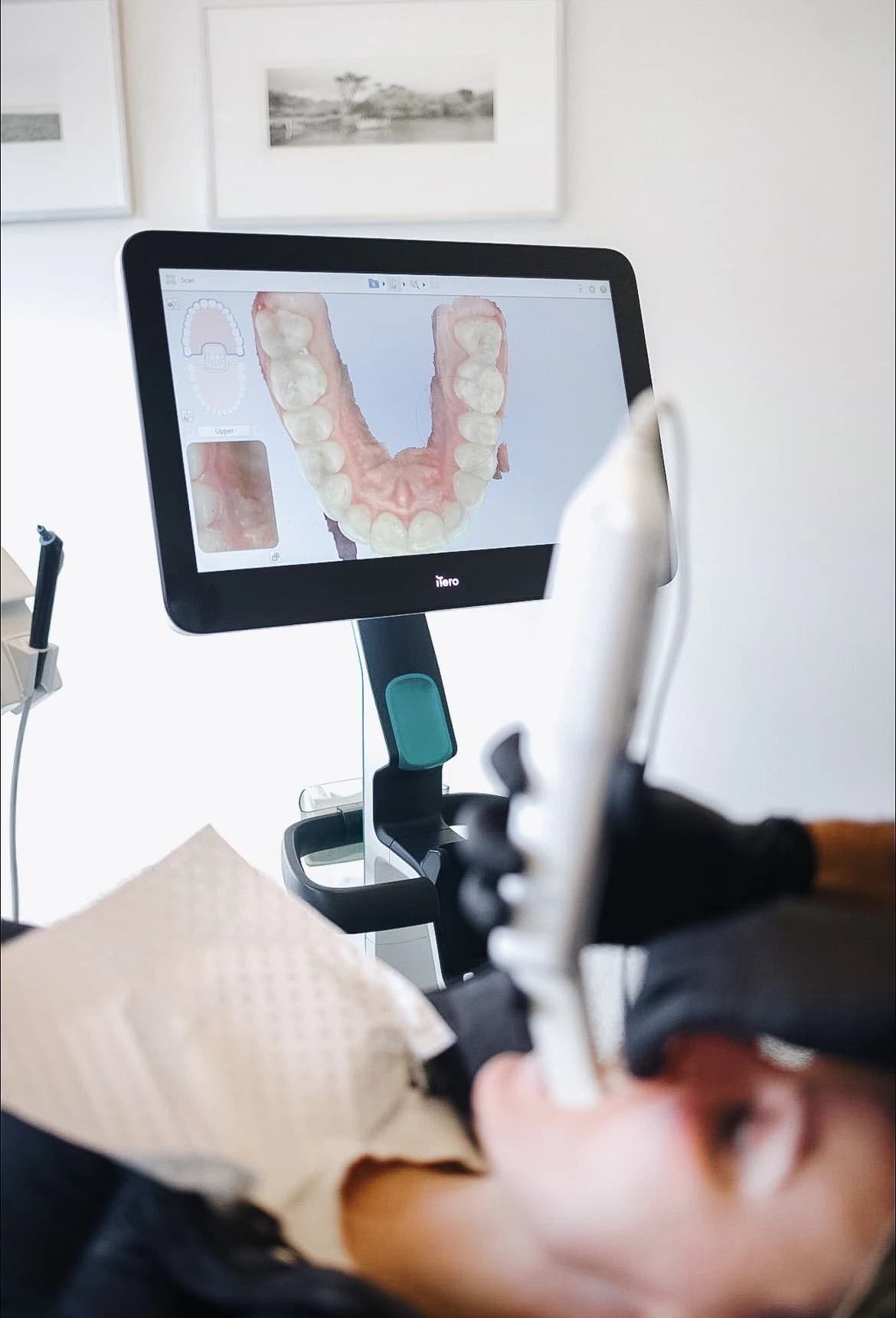
The first move is a free 3D CBCT scan, not another flat X-ray. A 2D film flattens everything into one plane and routinely hides the exact thing you're hunting for — a wisp of radiolucency at the implant apex, a lesion on the tooth next door, a fleck of cement, a fractured screw. CBCT shows the implant and everything around it in three dimensions, so the source of the drainage is something you can see rather than guess. When a tract can be traced, we sometimes thread a tiny gutta-percha point into it and scan again; on the image, that marker points straight back to the origin like a dye trail.
From there it's methodical. Percussion and mobility testing tell us whether the implant is still solidly integrated or has any give — a stable implant with a tract is a fundamentally more hopeful picture than a mobile one. We probe the pocket depth, check the adjacent teeth for their own pathology, and if the crown is cemented we look hard for retained cement. Dr. Sam Jain's background — a DMD with a master's in mechanical engineering — tends to show up here, because a lot of these late failures are mechanical stories: a screw that loosened and let the connection micro-move, a bite that overloaded one implant, a cantilever doing more work than it should. Reading the forces is half of reading the cause.
One quiet advantage in a case like this is continuity. When the same team plans, places, and restores an implant, the original 3D plan and the exact components used are on file — so if you're our patient, we already know the implant system, the abutment, the torque, and what the bone looked like on day one. Dr. Sam Jain and Dr. Arpana Gupta, both ICOI Masters, do all of that in-house rather than handing pieces to a fly-in surgeon, which is a big part of why patients choose us for exactly these puzzle cases. And if we didn't place it, the CBCT usually tells us most of what we'd otherwise have to look up.

What the Fix Looks Like — and It's Usually Not Removal
The treatment follows the cause, which is why the diagnosis has to come first. If the source is a neighboring tooth, we treat that tooth — often a root canal or, if it's cracked beyond saving, an extraction — and the tract next to the implant resolves on its own once the real infection is gone. If it's retained cement or a loose screw, the fix is often surprisingly small: clean out the cement, or remove, inspect, and re-torque a fresh abutment screw to spec. Dr. Arpana Gupta's training in endodontics is genuinely useful in this exact scenario, because telling apart a tooth-origin lesion from an implant-origin one is the kind of call that decides everything downstream.
When the source is a contained pocket of infection at the implant apex — retrograde peri-implantitis on an otherwise stable implant — the goal is to save the implant, not pull it. That can mean a small surgical cleanup: opening a flap, thoroughly disinfecting the lesion, sometimes removing the very tip of the implant (an implant apicoectomy), and grafting the defect so bone can rebuild. Published cases show integrated implants keeping their stability a year out after this kind of targeted rescue. It's more involved than a filling, but far less than starting over. If bone has been lost to true peri-implantitis, we add bone grafting to rebuild the foundation.
Removal is the last resort, reserved for an implant that's genuinely mobile or has lost too much surrounding bone to save. Even then it's not the end of the road — the site is cleaned, grafted, and re-implanted, and because our in-house zirconia mill turns around final teeth fast, you're not stuck in a temporary for months. The point of all this is simple: a small tract on a three-year-old implant deserves a real diagnosis before anyone reaches for the forceps. Most of the time there's a fixable cause hiding under that little bump, and finding it is exactly what a free consultation and 3D scan is for. If you're weighing options because an implant elsewhere is giving you trouble, our page on dental implants in Fremont covers how we approach both new cases and rescues.
Written and medically reviewed by Dr. Sam Jain, DMD, MS, and Dr. Arpana Gupta, DDS, MDS, who own this practice.
This article is patient education, reviewed by Dr. Sam Jain, DMD, MS, and Dr. Arpana Gupta, DDS, MDS, and is not a substitute for an exam. Treatment recommendations are made only after an in-person consultation and 3D imaging.